
Pin on Body (of) Work
Anatomy Explorer Abductor Digiti Minimi Muscle of Foot Abductor Hallucis Muscle Adductor Brevis Muscle Adductor Longus Muscle Adductor Magnus Muscle Biceps Femoris Muscle (Long Head) Biceps Femoris Muscle (Short Head) Calcaneal (Achilles) Tendon Dorsal Interosseous Muscles of Foot Extensor Digitorum Longus Muscle Extensor Hallucis Brevis Muscle
Tendon Diagram Leg / Cardiovascular System of the Leg and Foot nadilughaharabiahwall
It is made up of over 100 moving parts - bones, muscles, tendons, and ligaments designed to allow the foot to balance the body's weight on just two legs and support such diverse actions as running, jumping, climbing, and walking. Because they are so complicated, human feet can be especially prone to injury.
Foot and ankle anatomy, conditions and treatments
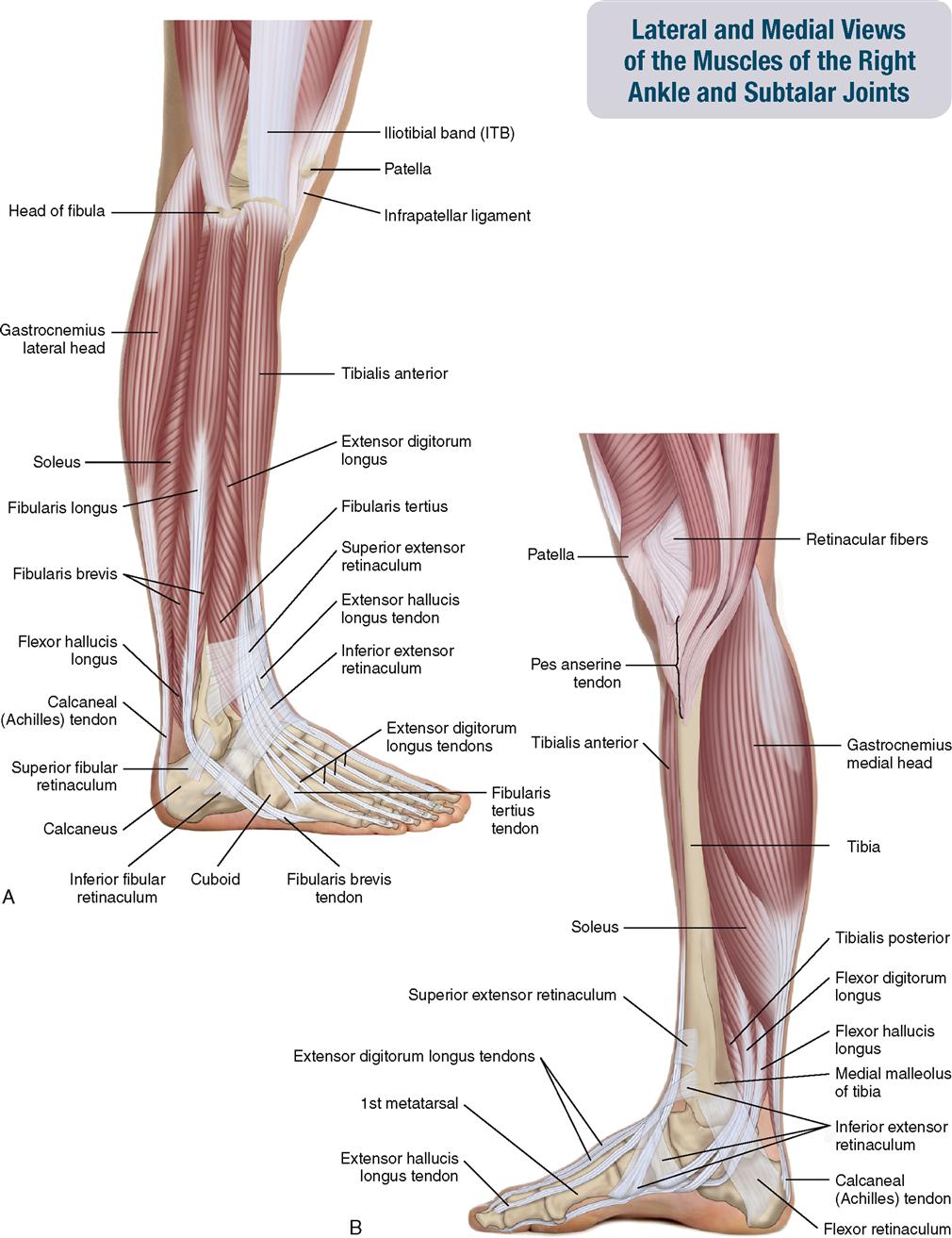
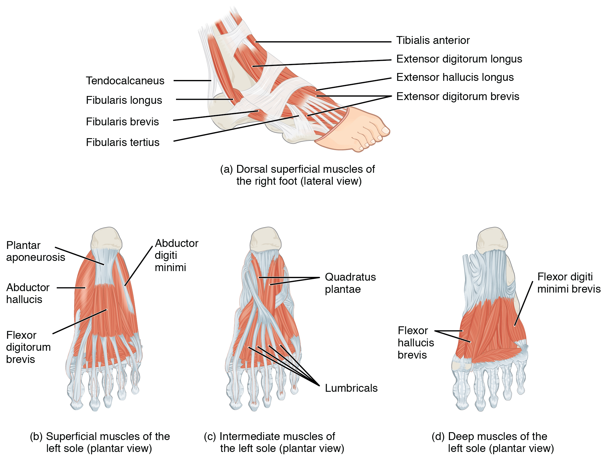
Introduction The foot is anatomically defined as the distal part of the lower extremity and encompasses all structures below the ankle joint. The muscles of the foot can be split into two groups, the extrinsic and intrinsic muscles. The extrinsic foot muscles are found in the lower leg and act to dorsiflex, plantarflex, invert and evert the foot.
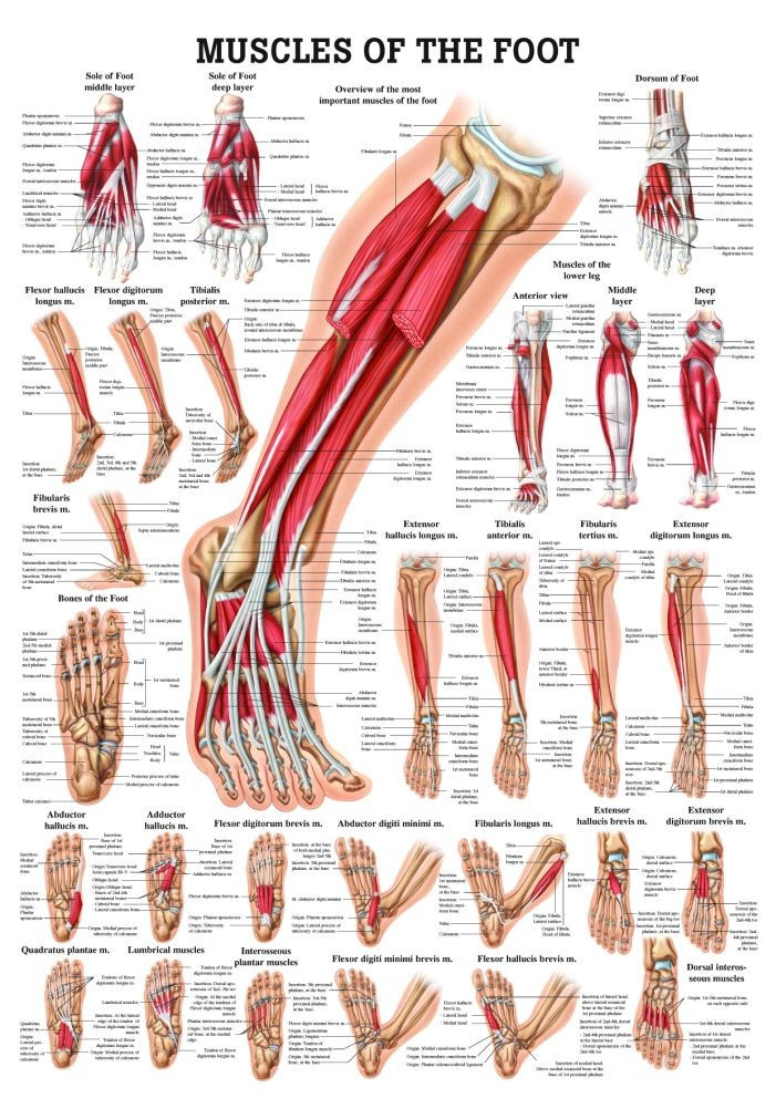
Human Muscles of the Foot Poster Clinical Charts and Supplies
Muscular System Muscles Muscles The 20-plus muscles in the foot help enable movement, while also giving the foot its shape. Like the fingers, the toes have flexor and extensor muscles.
Muscles that lift the Arches of the Feet
There are two intrinsic muscles located within the dorsum of the foot - the extensor digitorum brevis and extensor hallucis brevis. They assist the extrinsic muscles of the foot in extending the toes and are both innervated by the deep fibular nerve. Extensor Digitorum Brevis
11. Muscles of the Leg and Foot Musculoskeletal Key
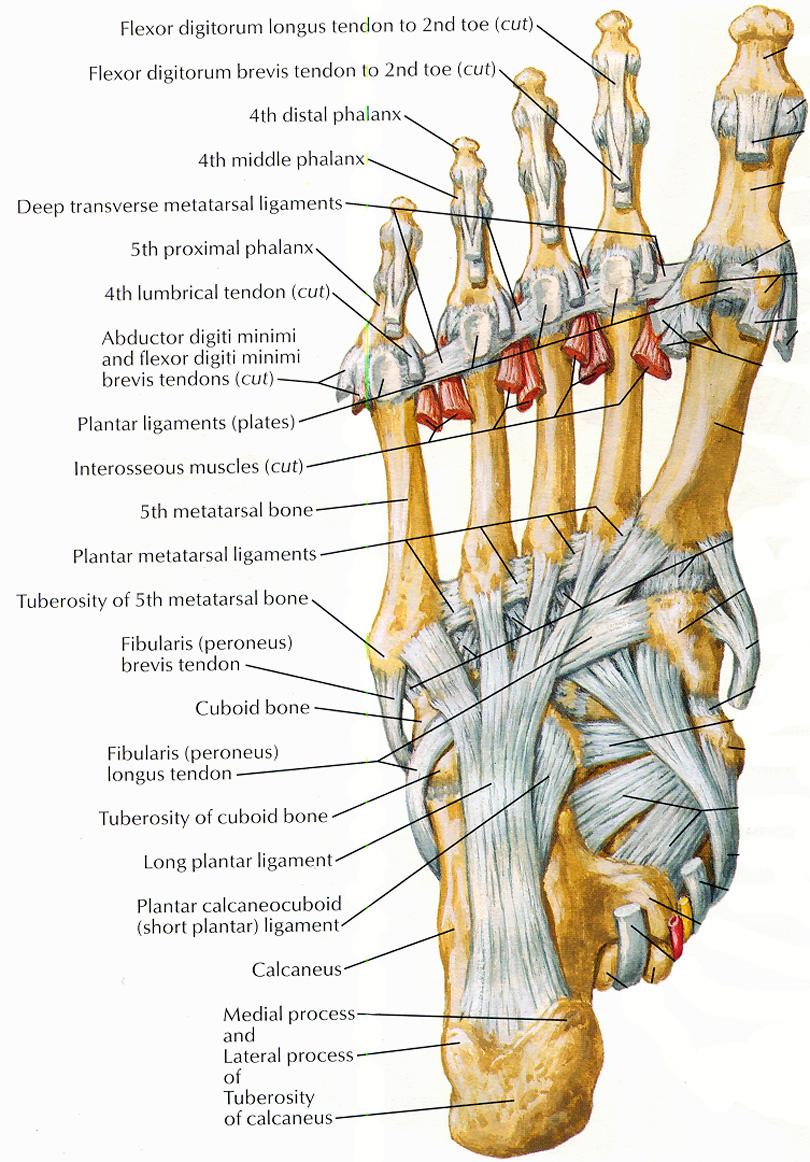
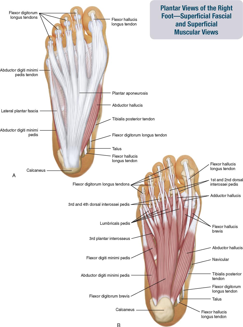
LABELED DIAGRAMS. Figure 1. Sections and Bones of the Foot A. Lateral (Left) B. Anterior (Right) Figure 2. Compartments of the Foot A. Cut Section through Mid-Foot. Figure 3. First Layer of the Foot A. Plantar View of Right Foot. Figure 4. Second Layer of the Foot A. Plantar View of Right Foot.
11. Muscles of the Leg and Foot Musculoskeletal Key
The Anatomy of Feet: Bones and Structure The foot is composed of 26 bones, making up about one-quarter of all the bones in the human body. These bones are divided into three main regions: the hindfoot, midfoot, and forefoot.

The extrinsic muscles that move the foot. Human muscle anatomy, Human body anatomy, Muscle anatomy
Anatomy and functions of the dorsal muscles of the foot shown with 3D model animation. The muscles of the dorsum of the foot are a group of two muscles, which together represent the dorsal foot musculature. They are named extensor digitorum brevis and extensor hallucis brevis . The muscles lie within a flat fascia on the dorsum of the foot.
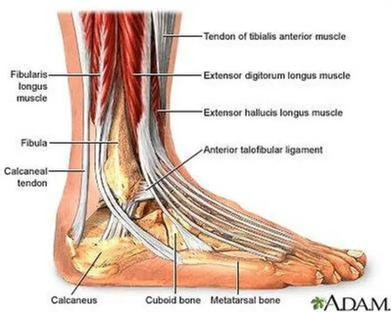
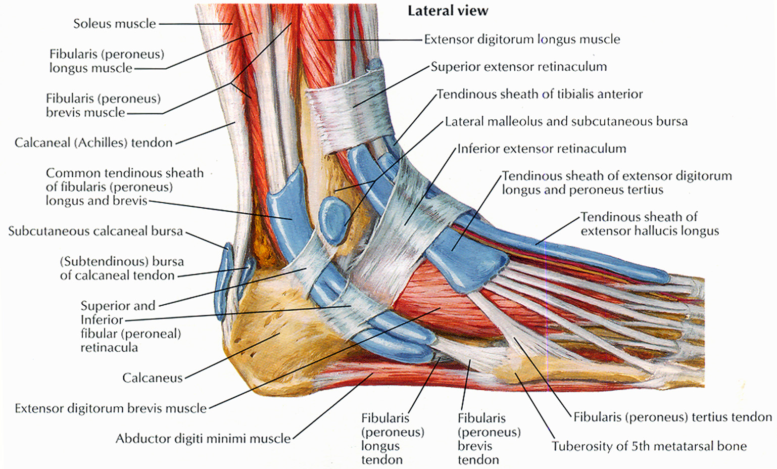
Pictures Of Ankle Muscles
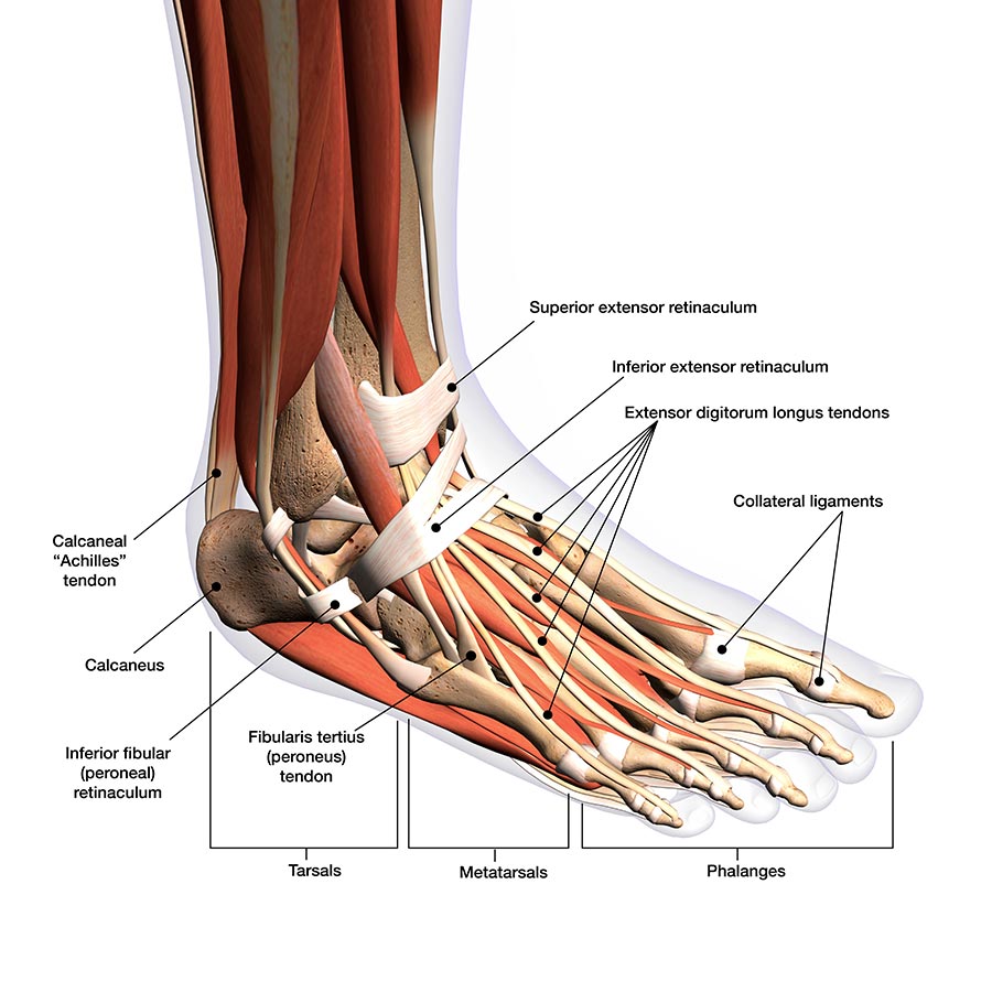
Ankle anatomy The ankle joint, also known as the talocrural joint, allows dorsiflexion and plantar flexion of the foot. It is made up of three joints: upper ankle joint (tibiotarsal), talocalcaneonavicular, and subtalar joints. The last two together are called the lower ankle joint.
Muscles that lift the Arches of the Feet
The foot is an intricate part of the body, consisting of 26 bones, 33 joints, 107 ligaments, and 19 muscles. Scientists group the bones of the foot into the phalanges, tarsal bones, and.

Foot Description, Drawings, Bones, & Facts Britannica
33 joints more than 100 muscles, tendons, and ligaments Bones of the foot The bones in the foot make up nearly 25% of the total bones in the body, and they help the foot withstand weight..
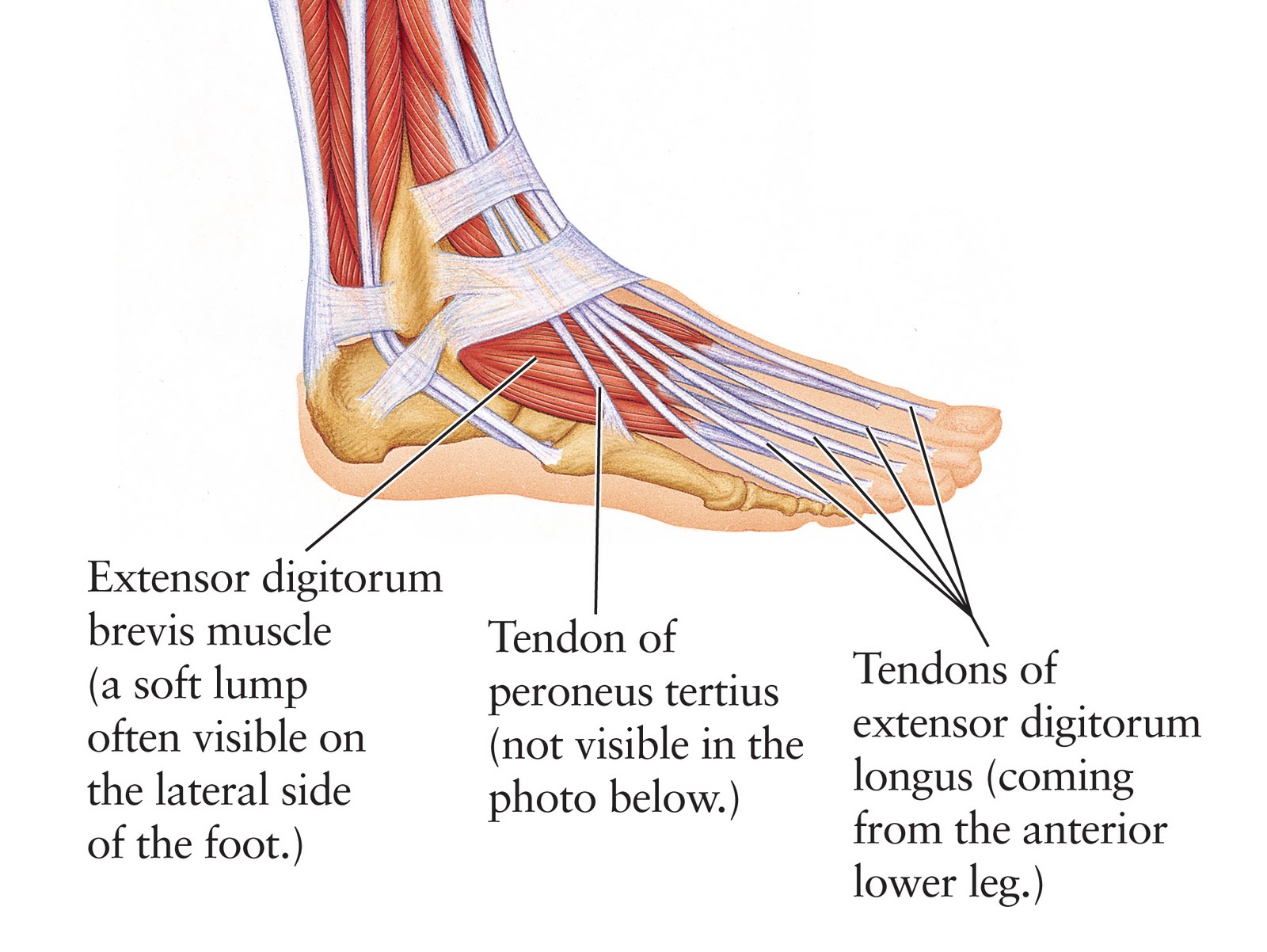
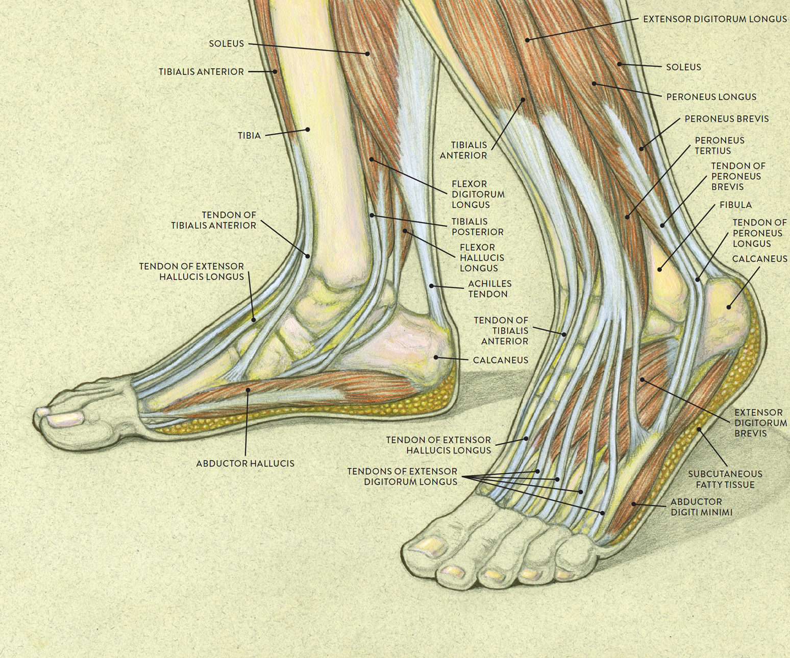
Human Anatomy for the Artist The Dorsal Foot How Do I Love Thee? Let Me Count Your Tendons
A foot pain diagram is a great tool to help you work out what is causing your ankle and foot pain. There are a whole range of structures e.g. bones, muscles, tendons and nerves which will each give slightly different foot pain symptoms.

Loading... Human anatomy chart, Foot anatomy, Nerve anatomy
The muscles of the foot are located mainly in the sole of the foot and divided into a central (medial) group and a group on either side (lateral). The muscles at the top of the foot fan out to supply the individual toes. The tendons in the foot are thick bands that connect muscles to bones.

Intrinsic muscles of the foot. Plantar intrinsics Layer 1 1 =... Download Scientific Diagram
Tibia Fibula Talus Cuneiforms Cuboid Navicular Many of the muscles that affect larger foot movements are located in the lower leg. However, the foot itself is a web of muscles that can.

Foot and Ankle Musculoskeletal Key
1. Foot Bones When thinking about foot and ankle anatomy, we usually divide the into three categories: the hindfoot, midfoot and forefoot. the hindfoot comprises of the ankle joint, found at the bottom of the leg. This is where the ends of the shin bones, the . Underneath this is the heel bone, aka the
Appendicular Muscles of the Pelvic Girdle and Lower Limbs · Anatomy and Physiology
It is a dorsiflexor of the ankle. Origin: Upper 1/2 of lateral and anterior surfaces of the tibia. Insertion: Inner surface of the medial cuneiform and 1st metatarsal. Actions: Inversion & Dorsiflexion. Innervation: Deep peroneal nerve. Daily uses: Walking - to lift the foot up and clear the ground.
