
Coils for MRI Questions and Answers in MRI
Radiofrequency (RF) coils are an essential MRI component used for transmission of the RF field to excite nuclear spins and for reception of the MRI signal. They play an important role in image quality in terms of signal-to-noise ratio, signal uniformity, and image resolution.
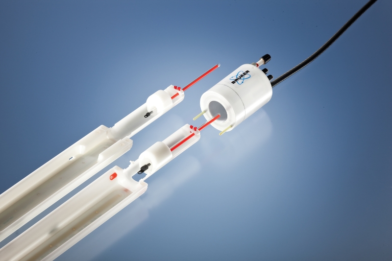
MRI RF Coil Imaging and Spectroscopy Bruker Bruker
Various radiofrequency (RF) ablation electrode designs have been developed to increase ablation volume. Multiple heating cycles and electrode positions are often required, thereby increasing treatment time. The objective of this study was to evaluate the performance of a high-frequency monopolar induction coil designed to produce large thermal lesions (>3cm) with a single electrode insertion.
4TIPS FOR EXTENDING THE LIFECYCLE OF MRI COILS
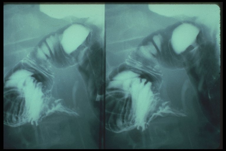
The coiled spring sign refers to the double contrast enema (DCE) appearance of appendiceal intussusception into the cecum, which is best seen en face. There is non-filling of the appendix with barium, and thin concentric rings of barium radiate outward from the appendix base, (Fig. 1) giving a spring-like appearance (Fig. 2 ).

GE Adaptive Image Receive Coil Shows Promise for WholeBrain Imaging
Radiofrequency (RF) coils are an essential MRI component used for transmission of the RF field to excite nuclear spins and for reception of the MRI signal. They play an important role in image quality in terms of signal-to-noise ratio, signal uniformity, and image resolution.
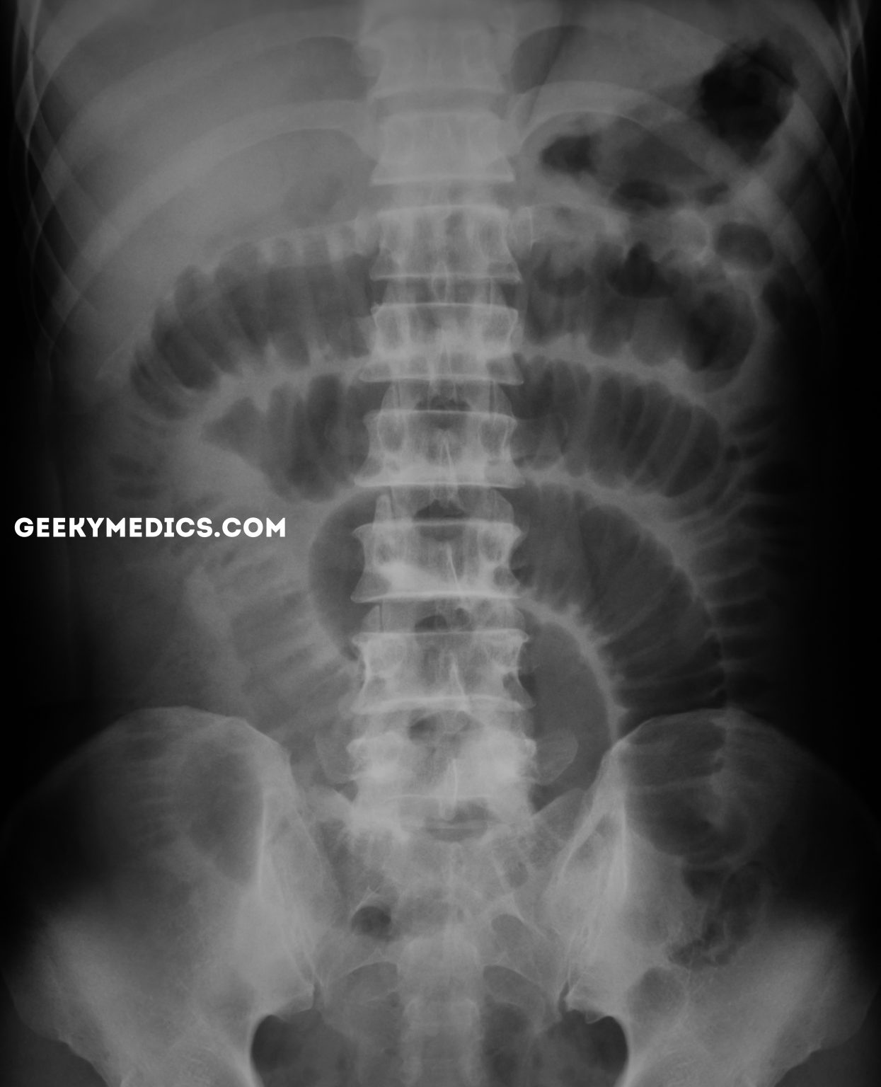
Abdominal Xray Interpretation
Coil-spring embolization is a procedure for treatment of pulmonary arteriovenous malformations. Herein is described a patient with hepatogenic pulmonary angiodysplasia ("pulmonary spiders") managed with this technique. Pulmonary angiodysplasia with hepatic cirrhosis is a well-described but poorly explained entity.

How DuoFLEX Flexible MRI Coils Boost MRI Scan Performance
The largest frequency shift and worst impedance matching were 3.6 MHz/−2.8 dB and 7.3 MHz/−3.2 dB for the conventional overlapped and self-decoupled coils, respectively. The normalized S21 of.

Spring ligament complex Illustrated normal anatomy and spectrum of
Coiled-spring sign of appendiceal intussusception Radiology. 1985 Apr;155 (1):41-4. doi: 10.1148/radiology.155.1.3975417. Authors M S Levine , S W Trenkner , H Herlinger , J D Mishkin , J C Reynolds PMID: 3975417 DOI: 10.1148/radiology.155.1.3975417 Abstract

All About Gradient Coils in Resonance Imaging (MRI
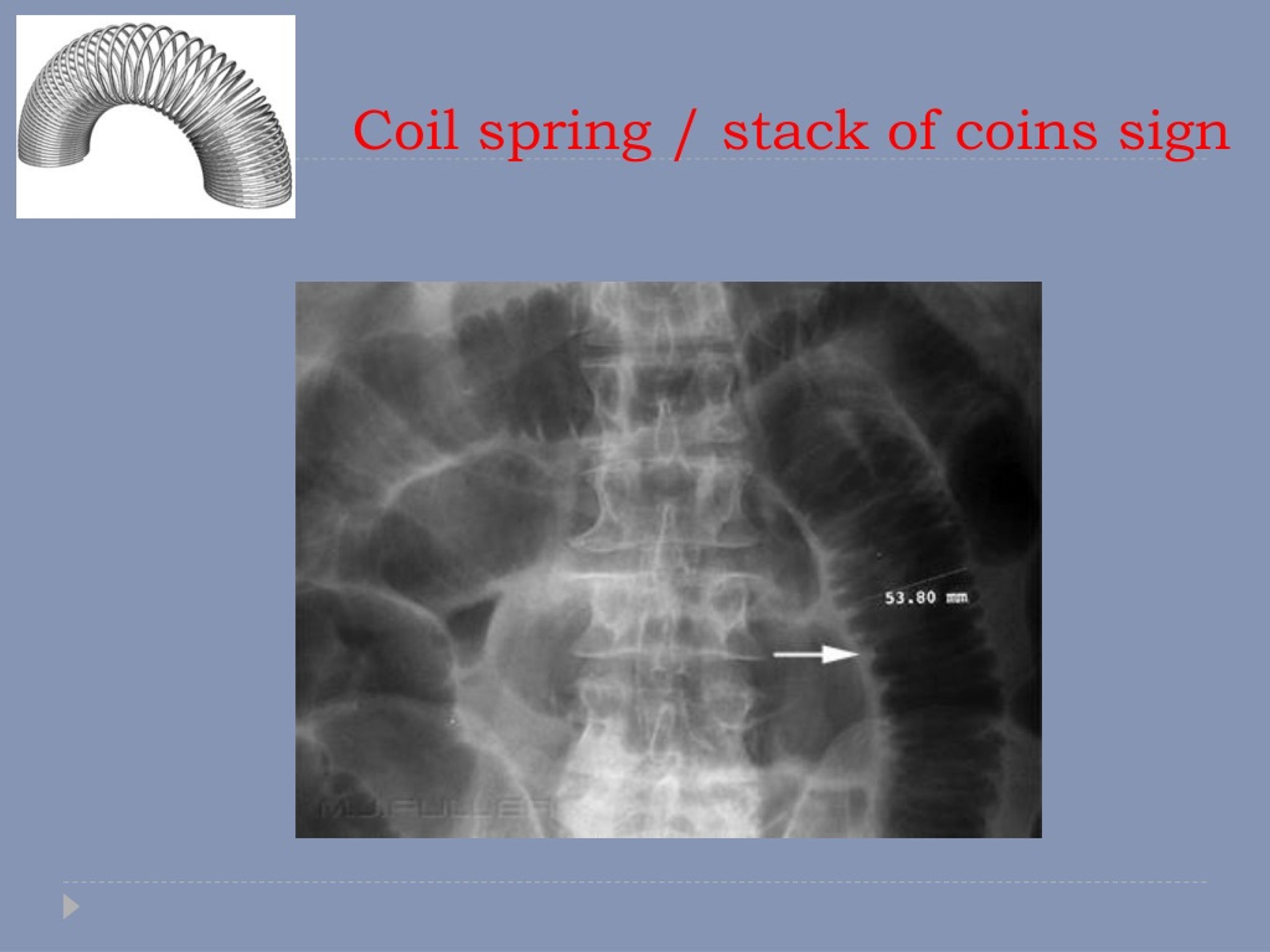
Coiled-spring appearance of Intussusception. Intestinal Intussusception in abdominal x-ray may produce the classic coiled-spring appearance (barium trapped between the intussusceptum and the surrounding portions of bowel). Note that Intestinal Intussusception is a major cause of small bowel obstruction in children (much less common in adults).

Spring ligament complex Illustrated normal anatomy and spectrum of
These scanners typically have spherical imaging volumes of 40-50 cm in diameter. With a subject present in the scanner, there is limited space for coil enclosures. RF coils must be in a protective housing for mechanical stability. These housings must be lightweight, non-magnetic, and non-conductive.
Integrated Coils Technology Resonance Imaging MRI
While the classic triad of intermittent abdominal pain, vomiting, and right upper quadrant mass, plus occult or gross blood on rectal examination, has great positive predictive value for intussusception in children 1, these findings, taken together, are seen in less than 20% of intussusception cases 2.

Spring ligament complex Illustrated normal anatomy and spectrum of
A biocompatible coil is one that is composed of primarily inert material that allows an effective treatment without the concern for a systemic host response. Metal alloys with a proved record for patient safety have been the main sources for coil production.
Many MRI coil failures are related to the cable Innovatus Imaging
The purpose of this study is to describe the sonography, CT, and MRI appearance of the Essure microinsert. Fig. 1 — Photograph of Essure microinsert (Conceptus, Inc.). Noted are two radiopaque markers at ends of inner (central) coil ( long arrows ), and two radiopaque markers at ends of outer (spring) coil ( short arrows ).
PPT Radiology of the abdomen PowerPoint Presentation, free download
The coils are made of soft platinum metal, and are shaped like a spring. These coils are very small and thin, ranging in size from about twice the width of a human hair to less than one hair's width. Healthcare providers also use coiling to treat a condition called arteriovenous malformation, or AVM.
coiled spring sign meddic
Coiled-spring sign of appendiceal intussusception. | Radiology Home Radiology Vol. 155, No. 1 Previous Next Coiled-spring sign of appendiceal intussusception. M S Levine , S W Trenkner , H Herlinger , J D Mishkin , J C Reynolds Published Online: Apr 1 1985 https://doi.org/10.1148/radiology.155.1.3975417 PDF Tools Share Abstract

Pin on Медицинская школа
In this image the intussusceptum (pink) is seen within the dilated intussuscipiens where a "stack of coins" or "coil spring effect" of telescoped valvulae are noted. 00512c01 small bowel intussusception upper GI UGI imaging radiology contrast X-Ray fx coil of springs stack of coins dx Peutz- Jeghers Courtesy Ashley Davidoff MD

MRI Options & Upgrades Coils
Given the growing variety of specialized coils available for neuroradiologic imaging applications, it is critical that radiologists use a coherent strategy for successfully matching these coils to specific imaging situations. First, fundamental concepts of coil design are reviewed.
