
How To Select The Right Tooth Pitch For Your Band Saw Blade SawbladeTV
The gull-wing appearance , also known as seagull erosions or sawtooth appearance , is classically seen in erosive osteoarthritis, typically on posteroanterior radiographs of the hands, although has also been reported in psoriatic and rheumatoid arthritis.

(A) Hyperkeratosis and irregular acanthosis with a sawtooth appearance
Saw-tooth appearance to the colon, usually sigmoid, with shortening of bowel, crowding of haustra and picket-fencing of folds Muscle spasm is present-may be relieved with glucagon Controversial as to whether this form can be symptomatic, i.e. pain Diverticulosis May be due to low roughage, high refined-fiber diet
Make a Saw Teeth at Home YouTube
Sawtooth pattern on ECG Last reviewed dd mmm yyyy. Last edited dd mmm yyyy Authoring team These include: if the P waves are about 300 per minute, and there is a 3:1 block, then the ECG will have a characteristic saw-tooth appearance

Saw Teeth Photograph by Jess Kraft Fine Art America
Appreciate the "saw-tooth" rectum. Maximal distention of the rectum is less than the sigmoid. Other things to consider when a newborn presents with dilated small bowel loops and a "low obstruction" are: meconium ileum, distal ileal atresia, colonic atresia, and small left colon syndrome.
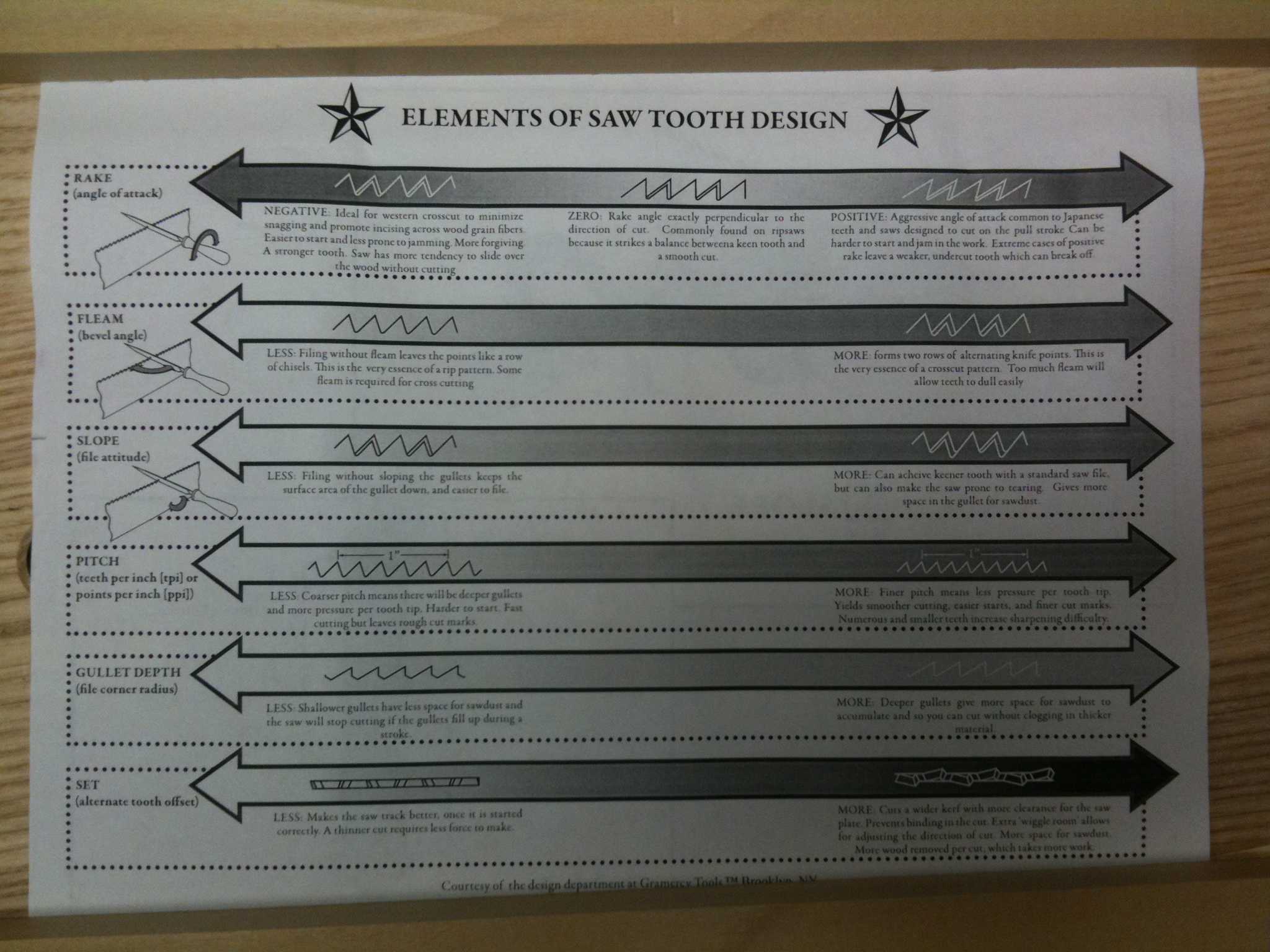
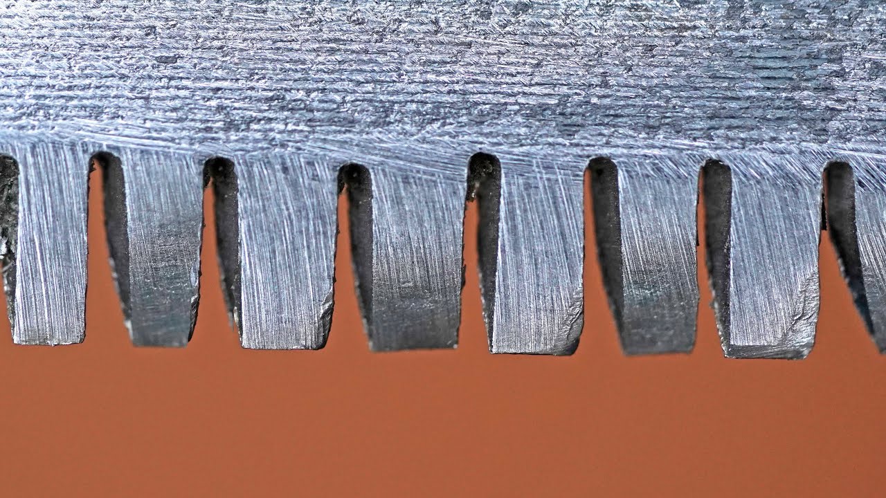
Understanding Saw Tooth Geometry The Renaissance Woodworker
The sawtooth wave (or saw wave) is a kind of non-sinusoidal waveform.It is so named based on its resemblance to the teeth of a plain-toothed saw with a zero rake angle.A single sawtooth, or an intermittently triggered sawtooth, is called a ramp waveform.. The convention is that a sawtooth wave ramps upward and then sharply drops.
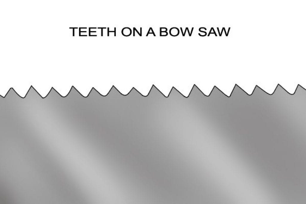
Crosscut Saw Teeth vs Rip Saw Teeth Wonkee Donkee Tools
Contrast enema with water soluble iodinated contrast demonstrated a transition zone in the distal descending colon with narrowed sigmoid and distal descending colon, "saw tooth" appearance of sigmoid colon and rounding of the flexures ( Fig. 2 a-c).
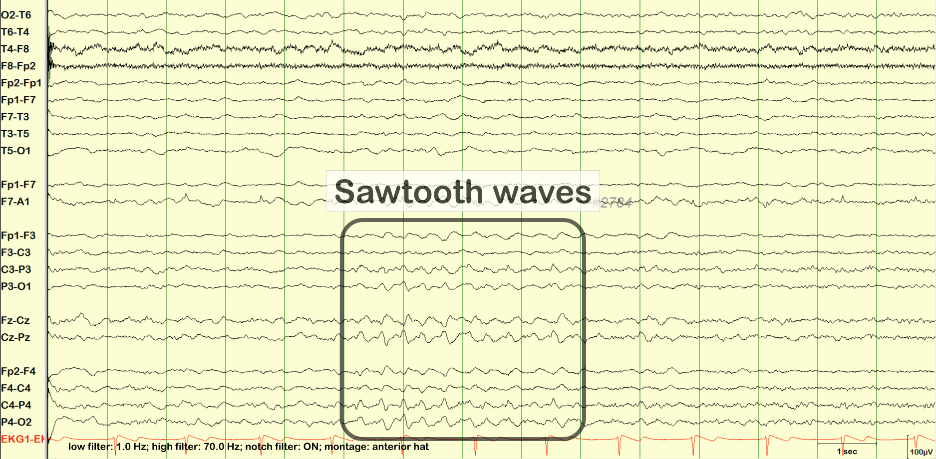
sawtooth waves
Saw tooth cardiomyopathy is an unusual and rare type of left ventricular dysplasia that is characterized by multiple projections of compacted myocardium that makes the appearance of 'saw tooth' in noninvasive imaging.

Antique Pruning Saw Restoration How To Make New Saw Teeth YouTube
Conversely, the lack of association of the radiologic findings of "sawtooth" appearance of mucosa and pneumatosis intestinalis is likely the result of there being a low incidence in this cohort. We found this somewhat surprising, given that when pneumatosis intestinalis or "sawtooth" appearance is present on imaging in a HSCR patient.

Crosscut Saw Teeth vs Rip Saw Teeth Wonkee Donkee Tools
Understand the concepts behind particular appearance and the derivation of the radiology sign - so that you never have to mug up
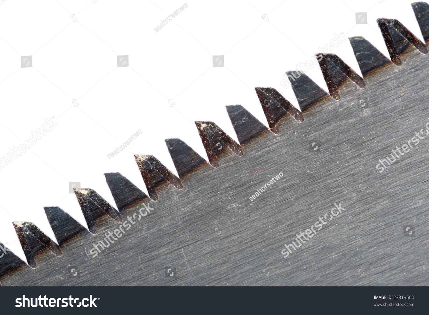
Saw Tooth. Isolated On White. Close Up. Stock Photo 23819500 Shutterstock
Download scientific diagram | (A) Hyperkeratosis and irregular acanthosis with a saw-tooth appearance and upper dermal band-like inflammatory infiltration (H&E, ×40). (B) Marked hyperkeratosis.

Free Image of teeth of a circular saw Freebie.Photography
Saw-tooth cardiomyopathy (STC) is a rare form of left ventricular (LV) dysplasia that may not be represented in the literature because it is underdiagnosed. Cardiac computed tomography is a valuable non-invasive study for the diagnosis of STC due to its capacity to rule out coronary lesions and identify this type of LV dysplasia.
Sharp metal saw blade teeth9733 Stockarch Free Stock Photos
DOI: 10.1007/s11325-016-1441-x Abstract Background: We retrospectively assessed the predictive value of the spirometric sawtooth sign in terms of the odds ratio (OR) of association with a diagnosis of OSA compared to those without the sign. Methods:

Circular Saw Blade Teeth Detail, Isolated Stock Photo Image of sharp
Barium enema demonstrates a reduced caliber rectum and sigmoid (the rectum is smaller than the descending colon) with a saw-tooth appearance to the wall. A transition point is seen at the junction between sigmoid and descending colon. 3 case questions available Annotated post evacuation film Fluoroscopy
Histopathology of lichen planus. Black solid arrow showing a saw tooth
The sacrum is recognizable as a horizontal hyperechoic curvilinear structure, and the L5 lamina has the typical "sawtooth" appearance. The structures of the vertebral canal are visible through the intervening gap. A distinguishing feature of the L5 lamina is its shorter superior-inferior width compared with the other lumbar vertebrae.

Free Image of wood saw teeth in closeup Freebie.Photography
Scanning power view of lichen planus shows a lichenoid reaction pattern (Figure 1) characterised by the combination of degeneration of the basal layer of the epidermis and a band like lymphocytic infiltrate obscuring the dermoepidermal junction. There is irregular epidermal hyperplasia forming a characteristic saw-tooth appearance with wedge.

Saw Teeth Close Up Stock Image Image 24266091
Saw-tooth cardiomyopathy has a characteristic appearance in CMR and/or echocardiography, which is distinct from LVNC. Natural history is unclear, as are potential complications, but conduction abnormalities are described in previously reported cases. The pathophysiology is unknown, but family screening and genetic analyses may be reasonable.
