PPT Review of Makhaarij PowerPoint Presentation, free download ID
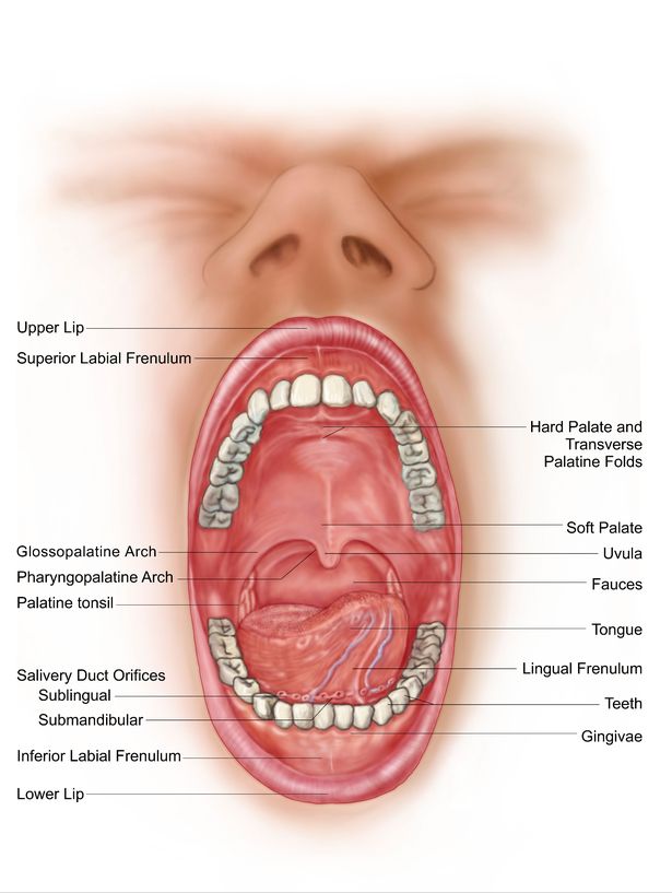
The Mouth The cheeks, tongue, and palate frame the mouth, which is also called the oral cavity (or buccal cavity). The structures of the mouth are illustrated in. At the entrance to the mouth are the lips, or labia (singular = labium). Their outer covering is skin, which transitions to a mucous membrane in the mouth proper.
Cancer of the Tongue, Mouth, Cheeks and Lips
A closed human mouth. The lips come together to close the opening of the mouth, forming a line between the upper and lower lip. In facial expression, this mouth line is iconically shaped like an up-open parabola in a smile, and like a down-open parabola in a frown.

Pin on Anatomy
The oral cavity, or more commonly known as the mouth or buccal cavity, serves as the first portion of the digestive system. It consists of several different anatomically different aspects that work together effectively and efficiently to perform several functions. These aspects include the lips, tongue, palate, and teeth. Although a small compartment, the oral cavity is a unique and complex.

Human Throat Anatomy Throat anatomy, Sore throat remedies for adults
Mouth Proper The mouth proper lies posteriorly to the vestibule. It is bordered by a roof, a floor, and the cheeks. The tongue fills a large proportion of the cavity of the mouth proper. Roof The roof of the mouth proper consists of the hard and soft palates. The hard palate is found anteriorly.

Diagram Showing Inside Of Mouth And Salivary Glands HighRes Vector
Browse 10,700+ human mouth anatomy stock photos and images available, or start a new search to explore more stock photos and images. Sort by: Most popular Anatomy of the mouth and tongue medical vector illustration on. Anatomy of the mouth and tongue medical vector illustration on white background eps 10
PPT Oral Cavity, Teeth, Tongue, and Salivary Glands PowerPoint
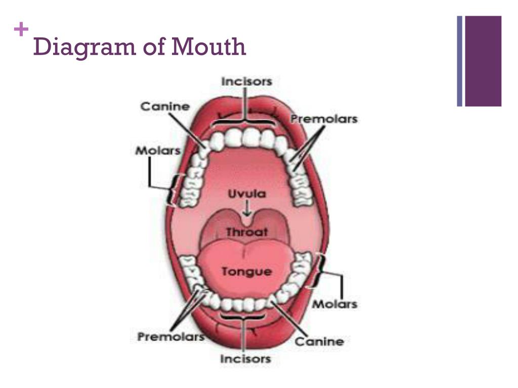
Mouth Mouth A molar tooth is located in the posterior (back) section of the mouth. It is found in most mammals that use their posterior teeth to grind food. Twelve molars are usually present.

fp protesis dental에 있는 핀
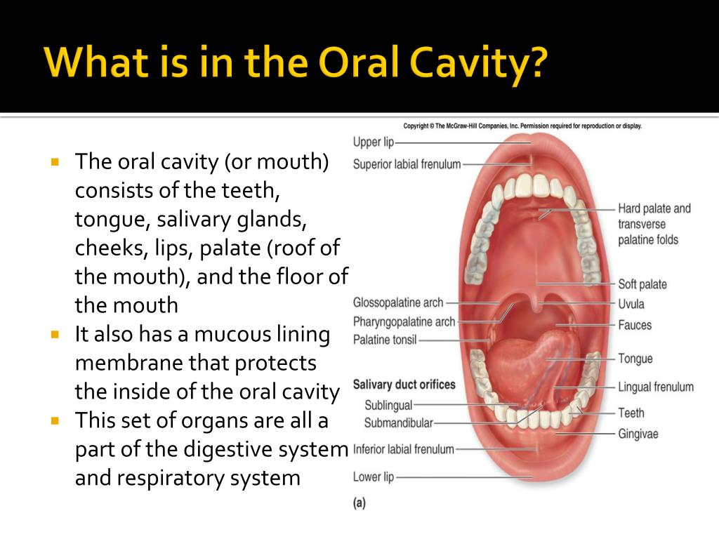
The mouth is also known as the buccal cavity or oral cavity. It includes the lips, cheeks and palate. It also encloses the tongue, teeth and salivary glands. The mouth anteriorly opens outside via the lips and posteriorly opens via the fauces (throat) into the pharynx.

Mouth Diagrams Printable 101 Diagrams
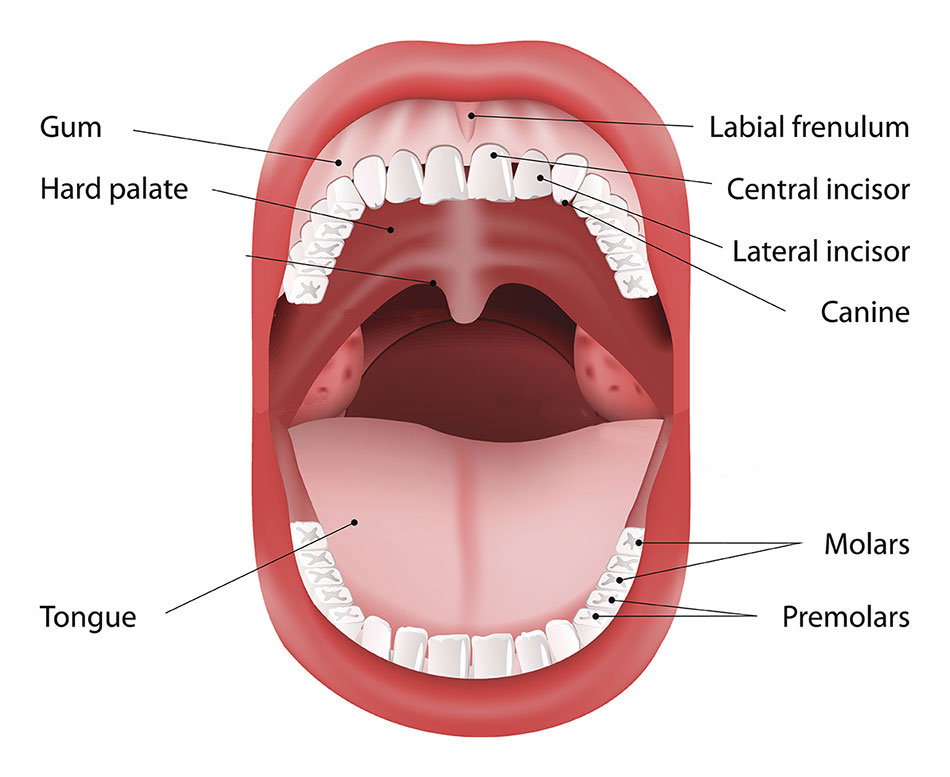
Anatomy of a Mouth. The mouth (oral cavity) consists of several components, including the teeth, gingiva (gums), tongue, palate, cheeks, lips and floor of the mouth. With the exception of the teeth, the mouth is lined by mucous membranes. The Teeth. The teeth are held within the jaw bones and serve several important functions beyond allowing.

Mouth Definition, Anatomy, & Function Britannica
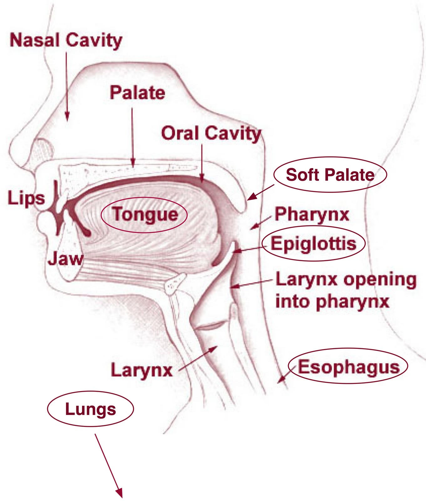
In this section, you will examine the anatomy and functions of the three main organs of the upper alimentary canal—the mouth, pharynx, and esophagus—as well as three associated accessory organs—the tongue, salivary glands, and teeth. The Mouth The cheeks, tongue, and palate frame the mouth, which is also called the oral cavity (or buccal cavity).

Mouth and Stomach Part 2 and 3 of the 5 Phases of Digestion
What's my mouth's function? Your mouth supports many daily functions, including: Breathing. Talking. Chewing. Tasting. Swallowing. Eating. Drinking. Mouth function in digestive system Your mouth is where digestion begins. When you chew food, your salivary glands make saliva (spit). Saliva helps break down starches in the foods you eat.
Medellitin The Taste Map, Umami and Kokumi In Taste)
What the inside of your mouth looks like with an intraoral camera. Roof of the mouth, teeth, gums, tongue, palate, bottom of the mouth.
Anatomy of your mouth and dental structure Dr Chauvin
This e-Anatomy module contains 110 illustrations on the oral cavity, the mouth, the tongue and the salivary glands. These fully annotated anatomical illustrations are presented as a comprehensive atlas of the oral cavity, specially designed for medical students, medicine residents and healthcare professionals. Material and methods

Diagram of the Mouth 101 Diagrams
The inside of the mouth is lined with mucous membranes. When healthy, the lining of the mouth (oral mucosa) ranges in color from reddish pink to gradations of brown or black.

FicheiroHead lateral mouth anatomy.jpg Wikipédia, a enciclopédia livre
The chief structures of the mouth are the teeth, which tear and grind ingested food into small pieces that are suitable for digestion; the tongue, which positions and mixes food and also carries sensory receptors for taste; and the palate, which separates the mouth from the nasal cavity, allowing separate passages for air and for food.

23.3 The Mouth, Pharynx, and Esophagus Anatomy & Physiology
The Mouth: Anatomy and 3D Illustrations The Mouth By: Tim Taylor Last Updated: Feb 16, 2022 2D Interactive NEW 3D Rotate and Zoom Anatomy Explorer Apex of Tongue Body of Tongue Epiglottis Esophagus Filiform Papillae Foliate Papillae Fungiform Papillae Gingiva (Gums) Glottis Hard Palate Lateral Glossoepiglottic Fold Lingual Glands Lingual Tonsils

Oral cavity anatomy with educational labeled structure vector illustration
The cavity is separated into anterior and posterior parts by the dental arches (or teeth): the anterior oral vestibule sits anteriorly to the teeth and behind the lips, whilst the oral cavity proper describes the area behind the teeth.
