The Larynx Anatomy and 3D Illustrations
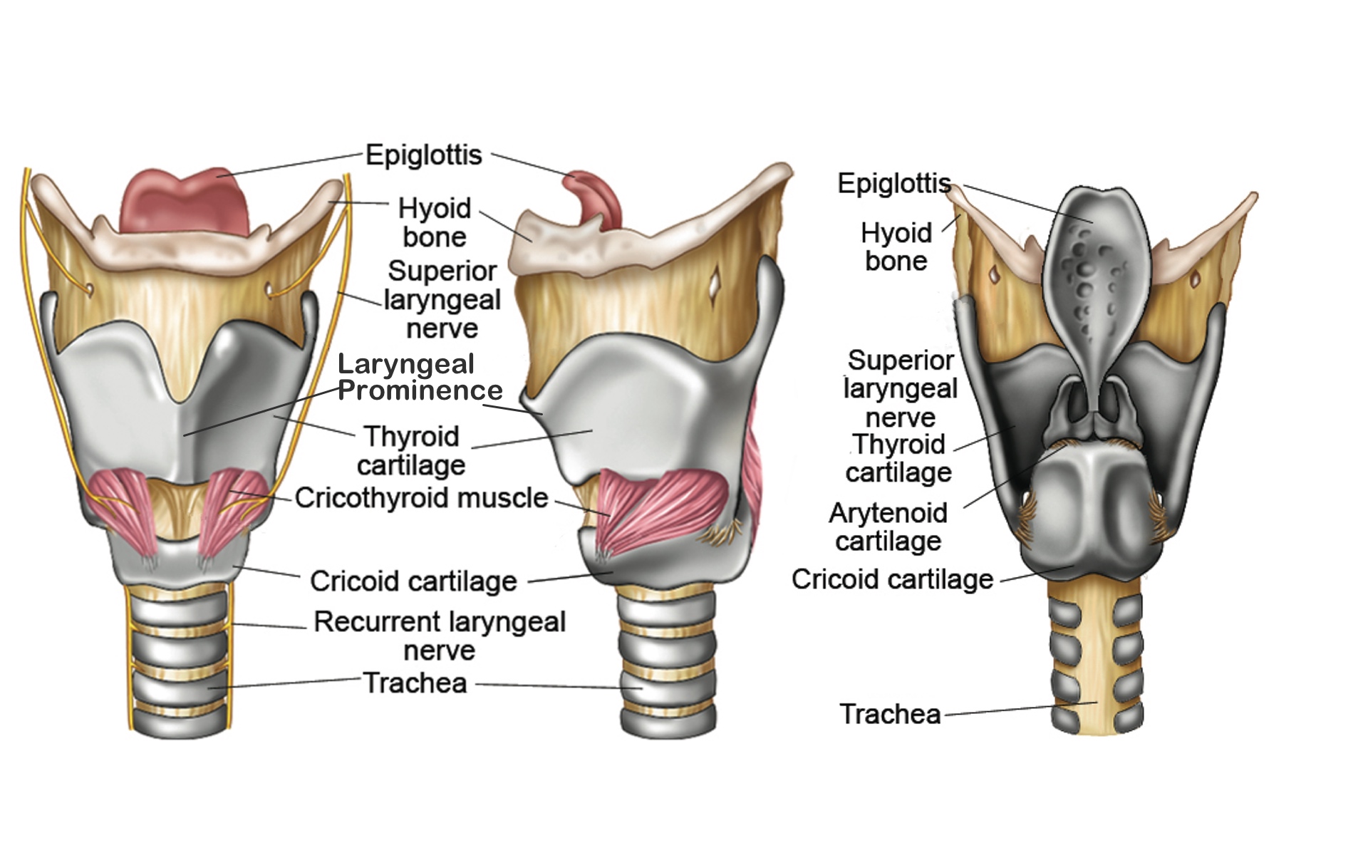
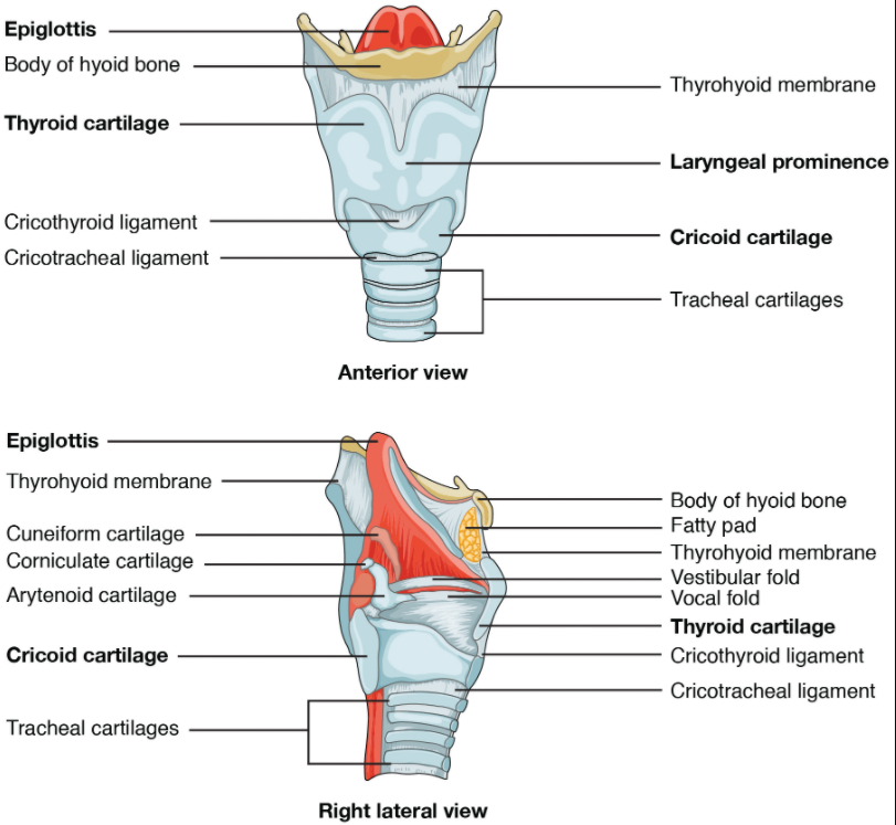
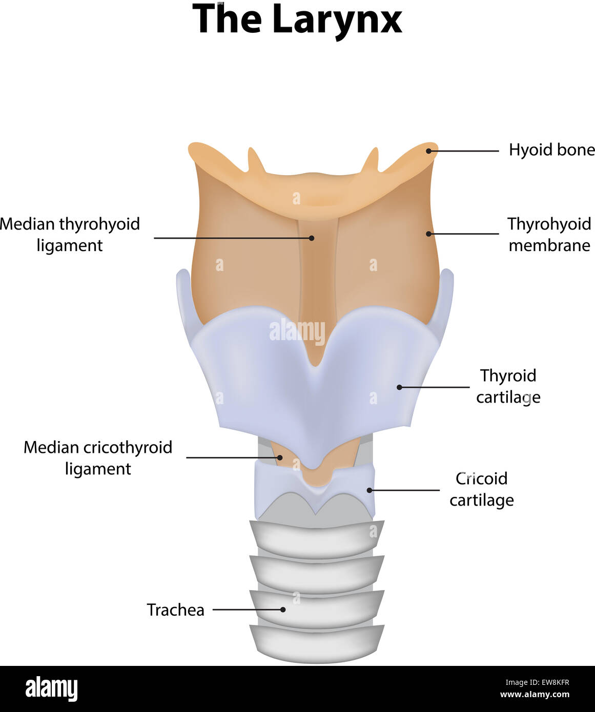
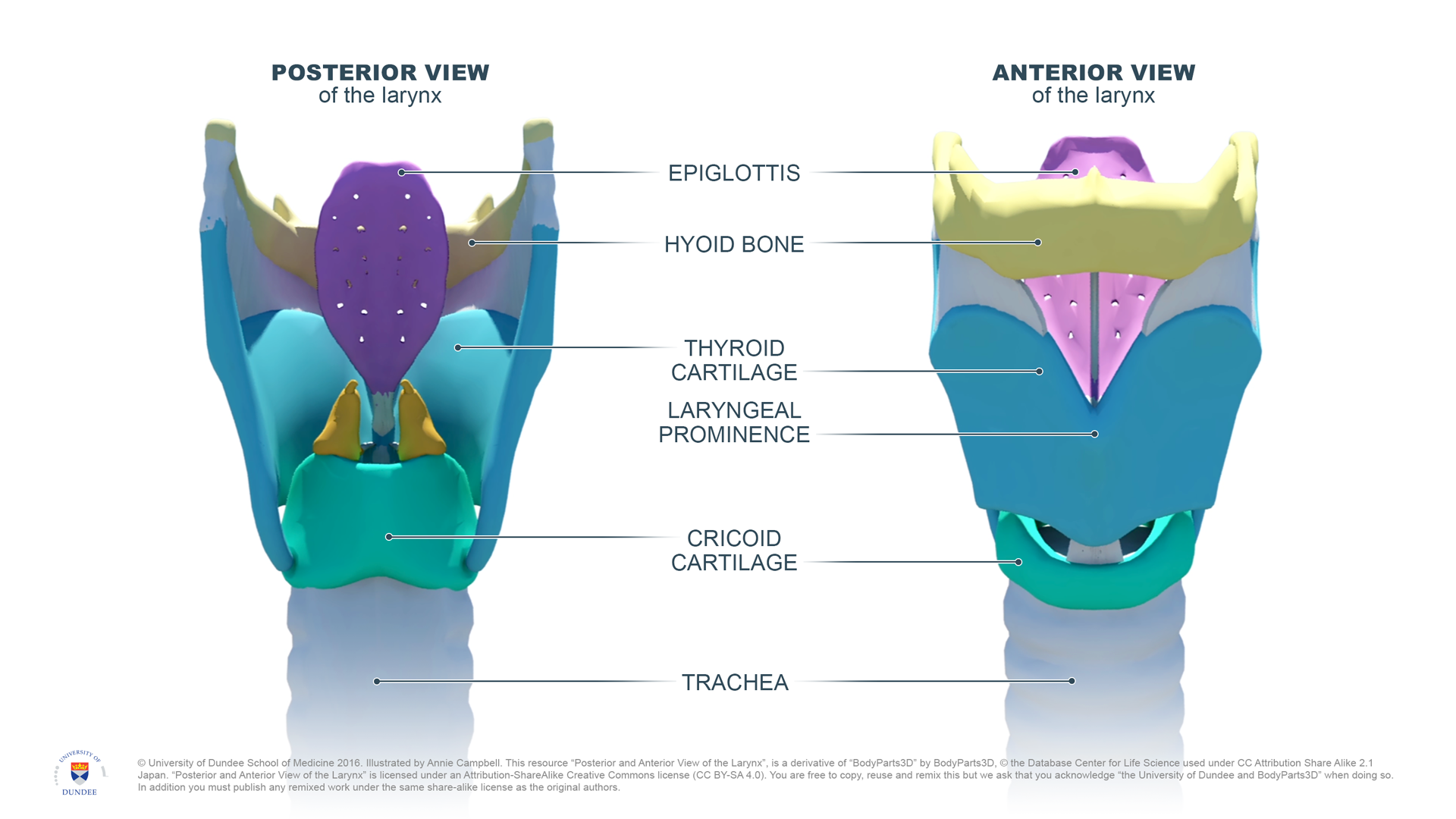
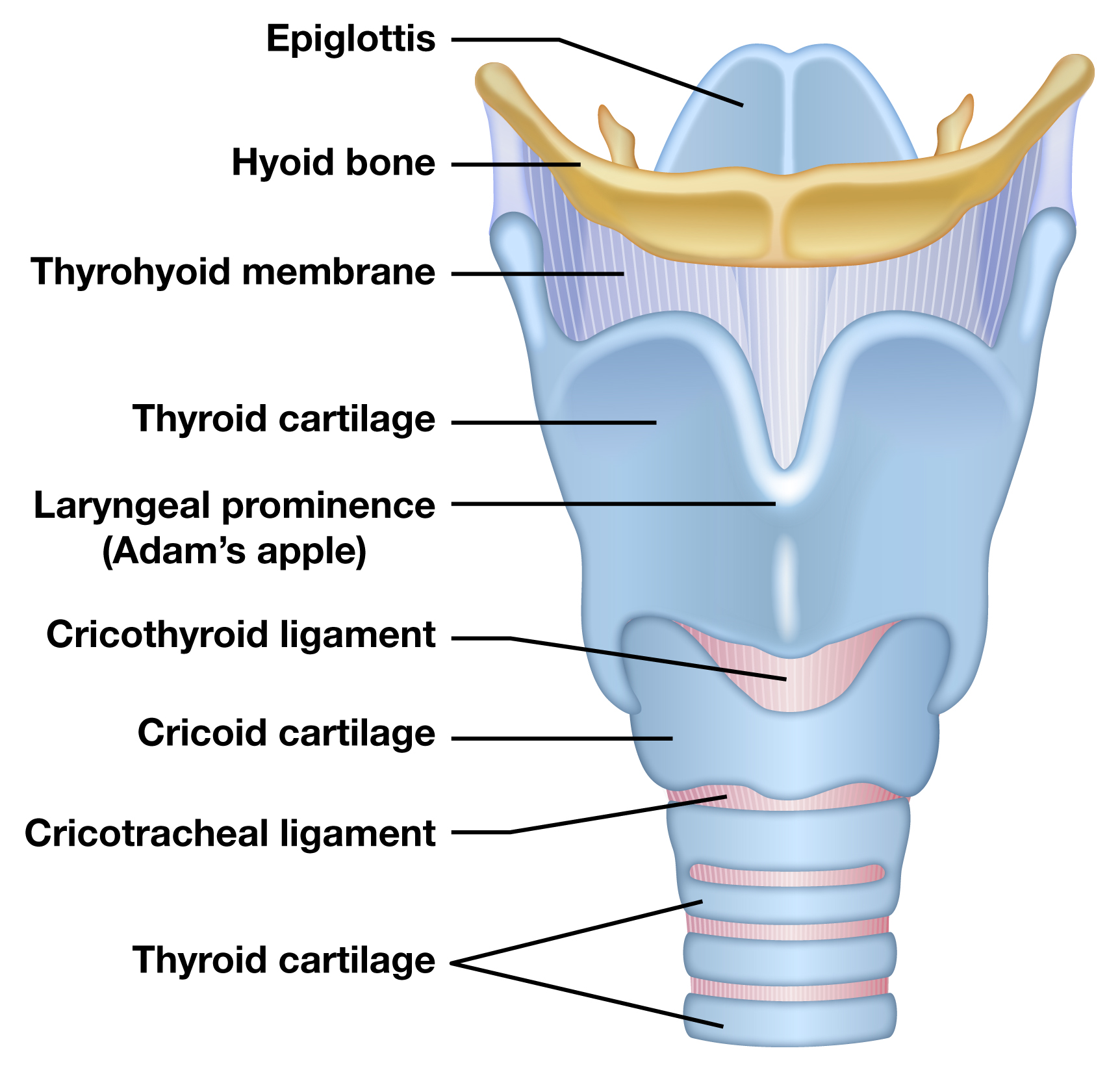
1/7 Synonyms: none The larynx is composed of three large unpaired cartilages (cricoid, thyroid, and epiglottis) and three paired smaller cartilages (arytenoid, corniculate, and cuneiform), making a total of nine individual cartilages. The thyroid cartilage is the largest of the laryngeal cartilages and is composed of hyaline cartilage.
The larynx home of the voice!
LARYNX AND TRACHEA. Click on a photo for a larger view of the model. Click on Label for the labeled model. Back to Respiratory System . Large Larynx (anterior) Large Larynx (posterior) Larynx (anterior) Label. Label: Label: Larynx (posterior) Larynx (sagittal cut) Trachea and Bronchi: Label:

10 best Larynx Model Project images on Pinterest Speech language
3D anatomy tutorial on the cartilages of the larynx from AnatomyZone For more videos, 3D models and notes visit: https://anatomyzone.comThis video is the fir.

Anatomy And Physiology, Community College, Slp, Image Shows, School
The larynx (voice box) is an organ located in the anterior neck. It is a component of the respiratory tract, and has several important functions, including phonation, the cough reflex, and protection of the lower respiratory tract. The muscles of the larynx can be divided into two groups; the external muscles and the internal muscles.
30 Label The Larynx Labels Design Ideas 2020
38.8k 67 Download 3D Model Triangles: 619.7k Vertices: 310.6k More model information Used as teaching material, this model depicts the muscles, cartilages and ligaments of the larynx. This was modelled utlising CT data, imported via Invesalius 3.1, and original sculpting in Pixologic ZBrush.
PHONATION Vocal Techniques for the Instrumentalist
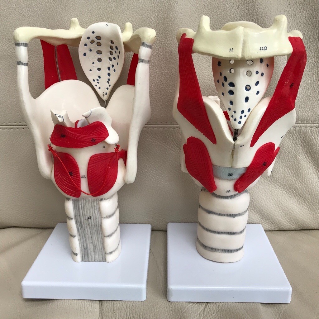
Product Group Summary Anatomical larynx models are ideal for active use in teaching and demonstrations. Views Of The Products Below
8.1 Organs and Structures of the Respiratory System Fundamentals of
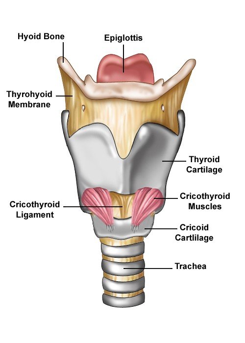
This functional larynx model shows the anatomy of the throat and the epiglottis, vocal cords and arytenoid cartilage are movable to demonstrate function. The following structures of the human larynx are also represented by this anatomical model: Hyoid bone. Cricoid cartilage. Thyroid cartilage. Thyroid.
Larynx Labeled Diagram Stock Photo, Royalty Free Image 84398827 Alamy
Anatomy of the Larynx - Download Free 3D model by University of Dundee School of Medicine (@tilt) [a00bc73] Connection error. Please try again. Anatomy of the Larynx 3D Model University of Dundee School of Medicine pro 5.7k 135.3k 251 Download 3D Model Triangles: 34.7k Vertices: 17.4k More model information
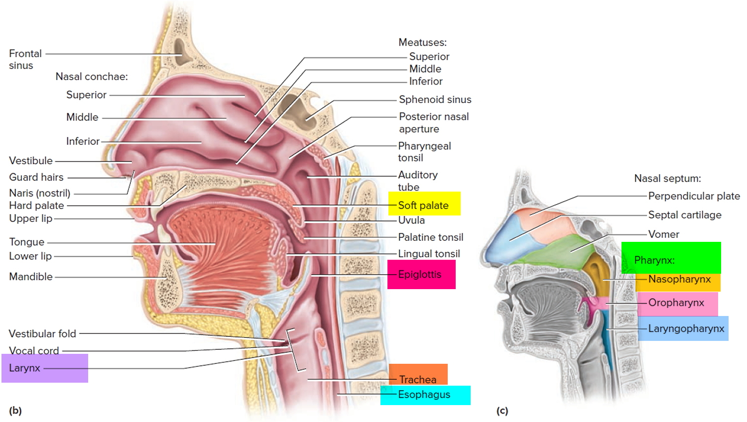
Pharynx Anatomy & Function in Respiratory System
Life Size Larynx Anatomical Model Anatomical Model Throat Anatomy Model Human Organs Teaching Prop Larynx Anatomical Model Anatomical Model Larynx $2214 3% off FREE delivery Nov 3 - 27

DIY Larynx Speech pathology grad school, Kids speech therapy, Speech
Currently, a static model is commonly used by speech pathologists to help educate their patients on larynx anatomy and physiology. The static model, although anatomically accurate, has limited capability in presenting larynx cartilages and muscle movements. This function is an integral component of patients' understanding of laryngeal function.
Anatomy Of The Larynx Anterior View slideshare
A prosection of a human larynx, pharynx, mandible, trachea, and esophagus. For more on the head and neck, visit: https://clinicalanatomy.ca/head.html Produced by the HIVE at the University of British Columbia. Credits: Dr. Claudia Krebs (Faculty Lead) Connor Dunne Ishan Dixit Monika Fejtek - Human Larynx - 3D model by UBC Medicine - Educational Media
Medical Images Art & Science Graphics
The model is specifically designed for easy display in classrooms and the doctor surgery. Our Anatomical Lung Model with Larynx can also be separated into removable parts, which allows for more anatomical accuracy and clearer representation. When the model is separated, it divides into a two-part larynx, trachea with bronchial tree, two-part.

The larynx (voice box). hyoid bone, laryngeal prominence, adam's apple
nid: 60506 Additional formats: None available Description: This model shows the anatomy of the larynx. Anatomical structures in item: Prominentia laryngea Cartilago thyroidea Larynx Os hyoideum Cartilago cricoidea Epiglottis Cartilago arytenoidea Cartilago corniculata Ligamentum vestibulare Ligamentum vocale Trachea Ligamentum hyoepiglotticum
Larynx Model Functional Human Larynx Anatomical Model approx 3x Life
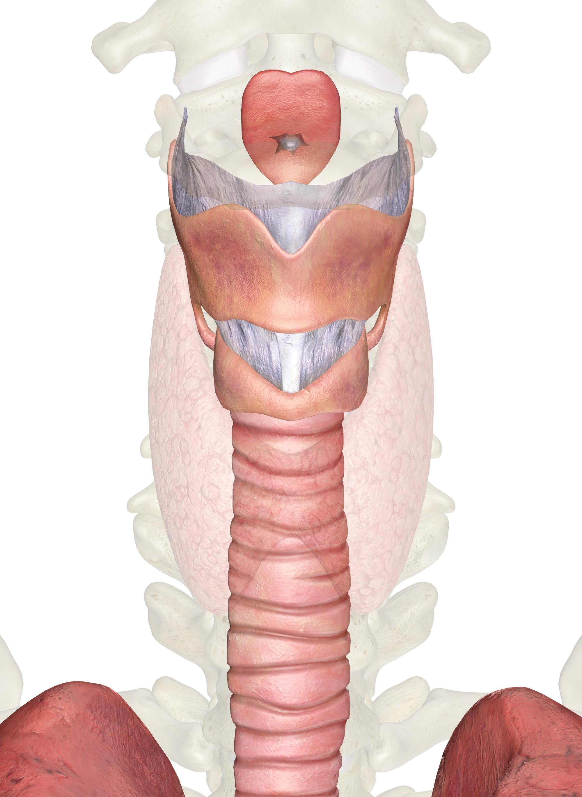
Your larynx is part of your respiratory system. It's a hollow tube that's about 4 to 5 centimeters (cm) in length and width. It lets air pass from your throat ( pharynx) to your trachea on the way to your lungs. Your larynx is also the reason you're able to make sounds, so it's often called your voice box. Advertisement.

The upper respiratory tract, nasopharynx, osopharynx, laryngopharynx
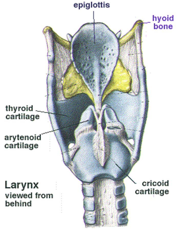
Structure and Function The larynx is a cartilaginous skeleton, some ligaments, and muscles that move and stabilize it and a mucous membrane. The laryngeal skeleton is nine cartilages: the thyroid cartilage, cricoid cartilage, epiglottis, arytenoid cartilages, corniculate cartilages, and cuneiform cartilages.

Schematic of the human larynx framework, based on Gray [6] a
This is the larynx model and will focus on the trachea, cricoid cartilage, false vocal cords, true vocal cords, epiglottis, glottis space, arytenoid cartilag.
